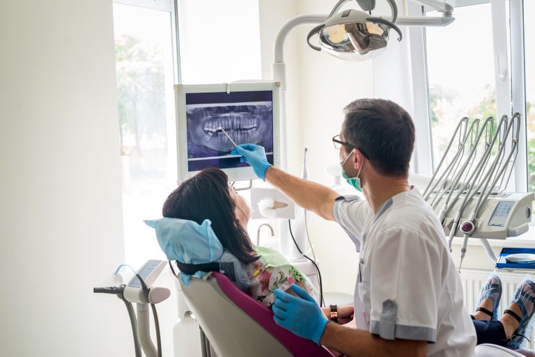
Annual x-ray imaging (radiographs) can be a polarizing topic in the dental office, as a patient or guardian of a patient it can be hard to determine the necessity. The answer in some cases may be, no you don’t need radiographs each year, maybe you need them every 18-24 months.

There are multiple factors that play a role in determining this; your current risk for developing decay, hygiene habits, previous history of decay, current medications, or even medical conditions. It is our job as clinicians to tailor the need for radiographs to each patient. That being said, a majority of individuals are at moderate-high risk for developing decay, which is why in most cases yearly radiographs are recommended.
Radiographs
Radiographs provide the dentist with information that cannot be obtained with a visual oral examination. Many times, cavities that appear in your general exam can only be seen on certain surfaces or teeth, or when they’ve already increased in size.
Our goal is always to catch decay as soon as possible. By treating decay in the early stages, we prevent further loss of tooth structure (weakening the tooth) as well as avoid more costly and invasive treatment. Most people would rather have a simple filling to treat early decay than a root canal and crown (or losing the tooth) because the decay has progressed.
Depending on your personal risk factors for decay, cavities can sometimes progress very rapidly, sometimes changing drastically within 12 months. In addition to identifying cavities, your dentist and hygienist use radiographs to evaluate bone levels.
Common Surface for Decay to Start
Radiographs allow us to look between the teeth, an area that cannot be seen in the oral examination and is the most common surface for decay to start. They also allow us to see the three layers of tooth structure (enamel, dentin, and pulp chamber) to determine how advanced a cavity has become.
When a cavity is still in the enamel layer, it will progress more slowly. When it surpasses the enamel and reaches the dentin layer of the tooth, it will be able to progress more quickly. The accelerated advancement at this stage is due to the softer structure of the dentin.
Even if a cavity does not infect the pulp chamber (the inner portion of the tooth containing nerves and blood vessels), a cavity in close proximity is sometimes enough irritation for the nerve of the tooth to become inflamed, causing irreversible pulpitis, typically resulting in a root canal.
Save Time, Money and Pain
Allowing your clinician to perform the radiographs they deem appropriate can often save you time, money and pain by finding cavities while they are less advanced. If you have concerns, please do not hesitate to discuss them with your hygienist. It is never our goal to expose anyone to unnecessary radiation.
In fact, all of our x-ray machines are completely digital (drastically decreasing radiation exposure) and lead aprons are available to anyone. We will discuss your particular risk factors and our recommendations for radiographs. You are ultimately in the driver’s seat and have every right to determine what treatment you do or don’t receive and we are willing to discuss this further at any of your visits.


